
The Basic Structure of Skin

Let’s talk the structure of skin and some science. It is such a surprisingly complex, sophisticated, clever organ!

A basic understanding of the skin is what drives all of the science and medicine behind the skincare that we recommend at Dr Emmaline’s Cosmetic Clinic.
The Epidermis
The top layer is the most superficial layer of the skin, known as the epidermis. It is extremely important from the point-of-view of an aesthetic doctor, as this is the layer that we see when we look at the skin. It is what gives the skin its unique texture and colour, and retains its moisture.
Keratinocytes are very important for your skin and are the most abundant cells in your epidermis. They’re found in all layers of the epidermis, changing shape as they migrate through each layer. They are produced by basal cells (stem cells) in the base of the epidermis at the dermal-epidermal junction. They produce keratin, an important structural protein that gives strength to your skin. As they move through the layers of the epidermis, they have unique characteristics, and they are what define the specific traits of each layer of the epidermis.
The epidermis consists of five sub-layers, the top layer being the stratum corneum. This is made of flattened (and dead!) keratinocytes stuck together with lipid-rich ceramides. These cells have lost their nucleus and granules. They create a watertight barrier for your skin. Skin is constantly being turned over or shed from this top layer, and the control of this is a very complex cell signalling process. When it goes wrong, you can get a number of skin conditions including psoriasis.
Underneath the stratum corneum, the thicker skin on the palms of your hands and soles of your feet have a clever extra layer called the stratum lucidem, adding a bit of protection against friction and other abrasive forces. These are tightly packed, transparent layers of dead cells.
Next is the stratum granulosum, a hydrophobic (water-repelling) layer that helps to waterproof your skin. This is the first living layer of your skin. Plastic surgeons are particularly interested in this layer as dermatohistopathologists will start measuring invasion of melanoma (skin cancer) from here! The flat cells here contain basophilic granules.
Then is the stratum spinosum, consisting of more keratinocytes linked together by desmosomes (“prickle cells”). The desmosomes are spikey in appearance - looking like spines sticking out of the cells - giving this layer its name. The desmosomes are complex molecules, important for cell adhesion (sticking the cells together). These cells are tightly packed to resist any stress and shearing forces. This layer gives the epidermis its strength.
Finally you have the deepest layer, the stratum basale. This is where a basal cell cancer arises. This is often only one cell thick, where keratinocytes divide to create new skin cells. This is a vitally important process, as it is responsible for maintaining the epidermis by constantly renewing it.
This deepest layer of the epidermis is also where you will find melanocytes, the cells that create melanin pigment which gives our individual skin its unique colour. Melanocytes can be activated by UV light. Melanin protects our skin from the sun by absorbing and scattering this light. This layer is also home to Langerhans cells, which act like macrophages (white blood cells) and have an immune function. In addition to this, you can find Merkel cells, which are involved in touch sensation, at this layer.

The normal cell cycle for the epidermis, also know as the process of keratinisation (describing the movement of the keratinocytes through these layers of the skin) lasts about 26-42 days. Again, as aesthetic doctors we care about this length of time because it means this is how usually long it will take before you start seeing the full effects of topical, medical-grade skin treatments. As we age, this time lengthens more and more as our skin is no longer quite as good at renewing itself. This is why skin starts to look duller.
The Dermis
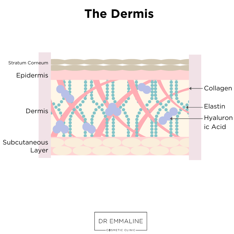
The dermis is the layer underneath the epidermis, lying between it and the subcutaneous fat. As we age, our dermis begins to thin and lose its moisture. This is the layer is stuffed with blood vessels, nerves, and sweat glands. The most important part of the dermis is arguably collagen. Collagen is an important protein, which is cross-linked into helices, and is the most abundant protein in our skin. When we are young, collagen fibres can glide against one another easily, giving smooth and flexible skin. As we age, we lose our collagen. This starts around the age of 25, and continues year after year! This is part of why aesthetic doctors are so obsessed with protecting your skin and collagen, even if you have naturally beautiful and clear skin. The idea is to preserve it while you still have it - you might not notice the difference today but you certainly will ten, fifteen, or twenty years down the line!
The dermis consists of two main parts - the papillary and reticular dermis.
The papillary dermis is the outer layer, filled with fibroblasts which produce dermal fibres. Fibroblasts are the most important cells in the dermis, producing all the proteins and enzymes in this layer. This layer also contains many capillaries, supplying nutrients to the dermis and the epidermis.
The reticular dermis is the thickest layer of the skin, and is full of blood vessels and elastin. It provides a very strong structure for the skin. Elastin is a very important part of our extracellular matrix. They exist in smaller numbers than collagen, and form a network around our collagen. This also gives skin its elastic property, something plastic surgeons are very familiar with and take into account when planning excisions or skin grafts.
Within the dermis, we also find mast cells. These calls release histamines, which we are most familiar with in hypersensitivity reactions and in causing an itching sensation.
In the dermis we also find sugars known as glycosaminoglycans (GAGs). They exist abundantly in the fibrous matrix and bind strongly to water. The most well known of these is a substance known as hyaluronic acid. This is a key ingredient in a lot of skincare products, and it is what most fillers are made of! Hyaluronic acid is famous for its ability to bind water, and gives the dermis its plump, full appearance. In young skin we see plenty of it at the interface of collagen and elastin fibers. However, as we age there is a distinct absence of hyaluronic acid!
The Hypodermis & Subcutaneous Layer
This layer is composed mainly of fat and collagen. However, again as we age we lose these layers of subcutaneous fat or they are migrated to other parts of the body. This is why another technique in aesthetic medicine is injected fat or filler into areas of natural fat pads to restore a youthful, full look. The hypodermis is stuffed full of fat cells, vessels, nerves, and connective tissue.
There you go - a brief whirlwind tour of the basic structure of our skin. Would you believe that this barely scratches this surface of what is happening in our skin? It helps us to appreciate the vast amount of medical knowledge that goes into treating the skin.


You might have heard buzz around the new vehicle taking the aesthetics and regenerative medicine world by storm. Exosomes are being touted as the “next big thing.” But what exactly are they, and what do we know about how they work?